Published Paper Review #2

Article: Eiji FurutaHiroshi OkudaAya Kobayashi, and Kounosuke Watabe*- Metabolic genes in cancer: their roles in tumor progression and clinical implications; Article accessed on 13th April, 2013

Metabolic Genes in Cancer

The equilibrium of energy homeostasis in a normal cell is due to three metabolic pathways which include glycolysis, lipogenesis and tricarboxylic acid (TCA) cycle which are in turn closely linked to amino acids. Cells use glucose as its main energy source. Glucose is converted to pyruvate through glycolysis whereas pyruvate is converted to acetyl-CoA which is then used in the TCA cycle in the mitochondria. ATP is generated by TCA via oxidative phosphroylation and citrate is transferred to the cytoplasm which is then converted to acetyl- CoA and used as a substrated for fatty acid generation via the lipogenesis pathway. Fatty acids are used as storage and also as a component in membrane biosynthesis as well as impacting cell signaling by protein modification.

Tumor cells are deemed hypoxic as they re-programme these metabolic pathways. Cancer cells have a high metabolism due to their active proliferation and motile nature. Based on studies and further research, it is believed that tumors rely on non-oxidative energy sources such as glyolysis and as a result tumor progression and tumorigenesis is due to the high number of genes involved in metabolic pathways.

In cancer cells, glucose uptake is significantly increased and oxidation phosphorylation in the mitochondria is significantly decreased. Due to the lack of oxygen and nutrition of the blood supply, rapidly growing cancer cells suffer, glucose metabolism and lactate generation is characteristic of tumor cells which results in hypoxia.

The link between glycolysis and tumorigenesis is a tumor suppressor, P53 which hypothetically blocks the glycolytic pathway through the tumor surpressor and induced glycolysis and apoptosis regulator. This occurs when the glycolytic metabolite fructose-2,6-bis-phosphate is decreased which induces glycolysis and inhibits gluconeogenesis.

Transporter proteins are used in the uptake of glucose across cell membranes. On these transporter proteins there are both amino and carboxy-terminal ends exposed to the cytoplasmic side of the plasma membrane. From the protein family, GLUT1 is found in several different ranges of cancers which include pancreatic, breast, brain and lung to name a few. It acts as an oncogene in cancers which can be mutated and found in high consistencies.

In tumor cells, the re-programming of the lypogenesis pathway is usually a significant alteration. Three genes participate in this process, ATP citrate lyase (ACLY), Acetyl-CoA carboxylase (ACC) and Fatty Acid Synthase (FAS). In FAS, pyruvate is converted to acetyl-CoA in the mitochondria and is then used in the TCA cycle.  Citrate is produced in the presence of sufficient amount of ATP and exported to the cytoplasm where it is catalyzed by ATP citrate lyase (ACLY). Cytosolic acetyl-CoA is then generated which is a key precursor of fatty acids. Acetyl-CoA is then carboxylated by ACC to synthesize malonyl-CoA which is then converted to palmitate (16-carbon saturated fatty acid) as the first fatty acid in lipogenesis by the key rate limiting enzyme.

Citrate is converted to Cytosolic acetyl-CoA by ACLY which is high in tumor cells. ACLY inhibitors block production of acetyl-CoA and consequently suppress cell growth in vitro and in vivo which results in the loss of tumorigenicity in vitro. It can therefore be determined that ACLY contributes to tumorigenesis and tumor cell survival.

The metabolic pathways of cancer cells are said to re-programme during tumors which is due to the alteration of metabolic genes. A high glucose uptake is observed in tumor cells whereas mitochondrial activity seems to be decreased in cancer cells.

Link: http://www.ncbi.nlm.nih.gov/pmc/articles/PMC2850259/

Published Paper Review #1

Article: T AlleyneI; S RoacheII; C ThomasI; A ShirleyIII; The Control of Hypertension by the use of Coconut Water and Mauby: Two Tropical Food Drinks; Article Accessed on 10th April, 2013

Care For A Drink, Anyone?

Did you know that coconut water and mauby are deemed two beneficial drinks to a healthy, “low-hypertensive” lifestyle?

Hypertension, as we all know is high blood pressure and is due to the elevation of arterial blood pressure which can have detrimental effects on the human body such as heart attacks, aneurysms, renal failure, to name a few. Hypertension is usually caused by smoking, obesity, lack of physical activity and diabetes, high sodium intake, stress or it may be genetically induced.

The Beginning

In the article, “The Control of Hypertension by the use of Coconut Water and Mauby: Two Tropical Food Drinks”, suggested that the consumption of these two drinks can gradually lower hypertension in the body. Various studies show that there is generally a low hypertension count in black populations of Sub-Saharan Africa, as compared to the Western Hemisphere, the USA and the Caribbean, where there is a high hypertension count.  Research has shown that black populations have a higher tendency in developing chronic high blood pressure and that symptoms develop at earlier stages in life.  Many treatments have been futile in attempting to cure this disorder, such as the beta blockers and ACE-Inhibitors. Over the centuries, this chronis disorder has been treated with the use of herbs and natural products, however with the emergence of modern medicines, these practices are now neglected.

Coconut water is used in many tropical countries as an ingredient in foods, and also as a beverage. The immature endosperm, a soft jelly is eaten whereas the dried endosperm is used in the making of pastries such as coconut tart. Cooking oil and margarine are two by-products of the dried endosperm. Based on scientific analysis, coconut water is high in sodium and potassium ions.

Mauby is manufactured from the bark extract of the mauby tree. It is a bitter, dark liquid in the concentrated form, and can be diluted with water and sweetened to taste. The concentrated form of mauby can be used to treat diabetes mellitus.

The Experiment

The study was conducted with the assistance of twenty-eight hypertensive individuals who were divided into four groups. Their systolic and diastolic blood pressures were observed for a period of two weeks before and after the, and once more for another two weeks while undergoing treatment. Group one which was the control, consumed drinking water, group two consumed coconut water, group three consumed mauby, and group four consumed a mixture of both coconut water and mauby.

Individuals studied were from two separate areas in Trinidad, West Indies. Twenty one individuals were from an industrial company in North-western side of the country and the remaining seven were from the St. Augustine Campus, UWI. The study was conducted based on the Declaration of Helsinki and Tokyo and was approved by the Ethics Committee of The University of the West Indies.

For each individual, all observations were recorded at approximately the same time of the day by the same researcher and they were asked to make no changes in their daily routines which ranged from eating habits to antihypertensive medication. The average blood pressures prior to and post the experiment were recoreded for each individual which included the included the highest and lowest values.

Assortment

The individuals were separated into their groups and each person was required to consume 300ml of the designated liquid at two points during the course of the day for the period allotted (two weeks). The careful preparation of the liquids to be consumed was carried out and dispensed into 300ml bottles.

What was the outcome?

For the controlled group, seven out of the eight individuals were found to have a high mean in systolic pressure. However there was no significant difference in the mean of diastolic pressure.

For the group who consumed only the coconut water, seven individuals experienced a decrease in mean of their systolic pressures. Two individuals had significant decreases. As compared to the systolic pressures, there were two cases with significant decrease in the mean of diastolic pressures.

For the group who received mauby, five out of seven individuals were able to complete the study. Two individuals experienced significant decreases in the mean of systolic pressures whereas the two other showed a general decrease and the remaining individual showed an increase in the mean systolic pressure.

Lastly, for the group who received the mixture of coconut water and mauby, there were high significant decreases in both the systolic and diastolic pressure averages as well as high decreases in the diastolic pressure averages.

Conclusion

Consumption of coconut water and mauby mixture resulted in the significant decrease in systolic and diastolic blood pressure averages as compared to the consumption of the separated beverages.

Link: http://caribbean.scielo.org/scielo.php?script=sci_arttext&pid=S0043-31442005000100002&lng=en

Video Review #2-Carbohydrates!

http://www.youtube.com/watch?v=Eqncc6u6Hms&list=PL3993356C72C83C43

Carbohydrates are organic molecules comprised of carbon atoms which are bound to other hydrogen and oxygen atoms. Carbohydrates can be divided into simple sugars and polysaccharides. They have several biochemical roles.

Firstly, they are energy sources, they allow the body to store energy in covalent bonds. For example C-C or C=O. Secondly, they are carbon skeletons which are able to build stuctures important in cell life. Carbohydrates are recognised by the formula (CH2O)n. For example, Hexose contains 6 carbon atoms therefore (CH2O)n would become C6H12O6.

Carbohydrates are divided into:
*Monosaccharides (1 Simple sugar)
*Disaccharides (2 Simple sugars)
*Oligosaccharides (3-20 Simple sugars)
*Polysaccharides (many Simple sugars)

Monosaccharides– 1 monomer include:
*Trioses (C3H6O3) which have 3 carbons, for example Glyceraldehyde and Dihydroxyacetone.
*Pentoses (C5H10O5)- example riboses-Alpha and Beta.
*Hexose such as Glucose (C6H12O6), Fructose, Glactose, Manose.

Disaccharides– 2 monomers bound by glycosidic bond formed by a dehydration reaction or condensation. When a glucose molecule binds to a fructose molecule, there will be a sucrose molecule developed. Lactose is formed by glucose and galactose. When two glucoses bind together, a maltose is formed.
Alpha linkages and beta linkages are found in disaccharides as well as polysaccharides. These glycosidic
linkages are the bonds between two simple sugars within a disaccharide or polysaccharide. Alpha
linkages are easily digested by the human body. Beta linkages are stronger than Alpha linkages
because they are more stable. Carbohydrates with Beta linkages are not easily digested by the
human body, except for lactose because most humans have an enzyme which breaks down this disaccharide.

Polysaccharides are large numbers of sugars joint together resulting in a polymer or macromolecule. There are several which are important to life such as celluose. Cellulose is the main component of the plant cell wall. It’s a linear glucose polysaccharide comprised of β (1–>4) bonds which link the glucose monomers to form fibres of great mechanical strength.

Starch, another essential polysaccharide, acts as the main form of storage for carbohydrates in plants. It is comprise of glucose molecules bound to one another. One of the main forms of starch is amylopectin which has ∝(1–>4)bonds and occasionally has ∝(1–>6) bonds which allow branching.

Glycogen is another essential polysaccharide, used by animal cells and human cells. It is the main form of storage of carbohydrates. Glucose is extracted whenever the body requires energy. This is stored as glycogen in the liver and muscles. Glucose is the monomer for the macromolecule glycogen. It is broken down and used as ATP which is used for metabolism in the body. Glycogen is highly branched.

I hope you enjoyed this video! 🙂 Here’s some facts about glycogen and starch.

A comparison of Starch and Glycogen:
Plants make starch and cellulose through the photosynthesis processes. Animals and human in turn eat plant materials and products. Digestion is a process of hydrolysis where the starch is broken ultimately into the various monosaccharides. A major product is of course glucose which can be used immediately for metabolism to make energy. The glucose that is not used immediately is converted in the liver and muscles into glycogen for storage by the process of glycogenesis. Any glucose in excess of the needs for energy and storage as glycogen is converted to fat.

Video Review #1-Nucleic Acids

This video, I found to be very educational. It’s done by one of three medical students who call themselves “The Salmonella Place”. They do videos based on several aspects of Medicine, Biochemistry being one of them.

The video begins with an introduction of nucleic acids, and their relation to DNA and RNA. A nucleic acid contains a chain of nucleotides linked together with covalent bonds to form a sugar-phosphate backbone with protruding nitrogenous bases. They are linear, biological molecules which are essential to life. The nucleotides are the basic unit of the nucleic acid. These molecules are able to store and express genetic information.

Next in the video, the tutor explains that nucleotides comprise of a nitrogenous Base, Pentose sugar and one to three Phosphates. Pentoses in the nucleic acid include Ribose and Deoxyribose which have bicarbons.

The base of the nucleotide is considered to be heterocyclic which simply means that there are two different rings of atoms- Nitrogen ring and Carbon ring. The base binds at position 1 or Carbon 1. The are two bases in nucleic acids. These are called Purines, which comprise of Adenine (A) and Guanine (G) and Pyridmidines which comprise of Cytosine (C), Thymine (T) and Uracil (U). Cytosine is found in both DNA and RNA, whereas Thymine is found only in DNA and Uracil, only in RNA. Between Adenine and Thymine there are 2 hydrogen bonds and between Guanine and Cytosine there are 3 hydrogen bonds. These are more heat resistant.

The phosphate groups are simply phosphate atoms surrounded by 3 hydroxyl (-OH) groups and an oxygen atom. Phosphate groups can bind both at the 3′ and 5′ position.

As compared to the nucelotide, the nucleoside lacks phosphate but comprises of only a nitrogenous base covalently attached to a pentose (ribose or deoxyribose). The formation of a nucleoside is due to the removal of the phosphate group of a nucleotide via hydrolysis.

He also spoke about a phosphodiester bond which is a covalent bond in RNA or DNA that holds a polynucleotide chain together by joining a phosphate group at position 5 in the pentose sugar of one nucleotide to the hydroxyl group at position 3 in the pentose sugar of the next nucleotide. This is called also a phosphodiester linkage.

Furthermore on nucleotides, they form ATP or Adenosine Triphosphate which is used for energy and storage. Also, they form the Cyclic Adenosine Monophosphate or CAMP which is used for regulatory functions.

Lastly, nucleotides are named after the base component. For example, with the base Adenine, the nuceloside will be Adenosine, Thymine, would be Thymidine. When a nucleotide is named after the base, it is also named after the number of phosphate groups. For example, a nucleoside with 1 phosphate group is called Monophophate such as AMP- Adenosine Monophosphate.

This video was found to be brief and informative, and I do hope you enjoy! 😀

Multiple Choice Questions On Glycolysis

Image

Multiple Choice Questions for Glycolysis

1) The second priming reaction in glycolysis is catalyzed by which enzyme?
a. Phosphohexose Isomerase
b. Aldolase
c. Pyruvate Kinase
d. Phosphofructokinase-1
e. Glyceraldehyde 3-Phosphate Dehydrogenase

2) When pyruvate is converted to Acetyl-CoA, the enzyme which catalyzes this reaction is
a. Hexokinase
b. Pyruvate Kinase
c. Phosphoglycerate Kinase
d. Phosphofructokinase-1
e. Pyruvate Dehydrogenase Complex

3) The enzyme which is responsible for the splitting of glucose molecules in glycolysis is
a. Hexokinase
b. Aldolase
c. Enolase
d. Phosphoglycerate Mutase
e. Phosphohexose Isomerase

4) When Dihydroxyacetone Phosphate is converted to Glyceraldehyde 3-Phosphate, the reaction is catalyzed by
a. Enolase
b. Phosphoglycerate Mutase
c. Triose Phosphate Isomerase
d. Glyceraldehyde 3-Phosphate Dehydrogenase
e. Pyruvate Kinase

5) Which reaction is considered the most energetically favorable reaction in glycolysis?
a. PEP converted to Pyruvate, catalyzed by Pyruvate Kinase
b. 2-Phosphoglycerate converted to PEP, catalyzed by Enolase
c. Fructose 1,6 Bisphosphate converted to G3P and DHAP, catalyzed by Aldolase
d. Glucose to Glucose 6-Phosphate, catalyzed by Hexokinase
e. G3P to 1,3 BPG, catalyzed by Glyceraldehyde 3-Phosphate Dehydrogenase

Enzymesssss

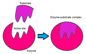
Enzymes are biological catalysts which speed up all processes via a chemical reaction by providing an alternative pathway with lower activation energy. Most enzymes are proteins (but not all proteins are enzymes) and some are RNA molecules. They are large molecules which consist of amino acids however, only a small portion of the enzymes play a role in the catalysis of the biochemical reactions. This portion is called the active site. The enzymes act upon molecules called substrates and this reaction occurs in the active site. Hence, an enzyme-substrate complex is formed. This bonding is due to hydrogen and hydrophobic bonds.

E + S ES → E + P

E= Enzyme
S= Substrate
P= Product

enzym

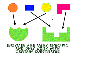
Enzymes work to lower the activation energy. They are specific and can only work with certain substrates. Denaturation of an enzyme can occur due to high temperatures and pH values, however when an enzyme becomes denatured the shape of the active site is altered and the substrate molecule is no longer able to fit. The activation energy is the minimum amount of energy required for the reaction to occur. When the activation energy is lowered, the more substrate molecules will be converted to product per unit time, hence there is more energy and the reaction occurs at a faster rate.

When an enzyme and substrate bind at the active site, a process called catalysis occurs. Catalysis occurs when the substrate is altered and can be broken down or bound to another molecule. This allows for the creation of a new molecule. The enzyme then releases the substrate and returns to its normal state however, the initial molecule has been modified. It is now called the product.

spec1

spec

Enzymes exhibit several degrees of specifity. These include:


  • Relative Specificity– where there is an interaction of a molecule with several substrates but with different affinities;
  • Absolute Specificity– refers to the situation whereby the enzyme catalyzes one reaction;
  • Group Specificity– occurs when the enzyme acts only on molecules with specific functional groups, for example, the amino and phophate groups;
  • Linkage Specificity– occurs when the enzyme acts on a specific type of chemical bond despite the molecular structure;
  • Stereochemical Specificity– occurs when the enzyme is acting on a steric or optical isomer.

Enzyme regulation occurs due to inhibitors. These inhibitors are molecules which are used to alter the catalytic reactions occurring, thus slowing down the reaction. Inhibitors bind reversibly or irreversibly. Reversible inhibitors are bound non-covalently and can be detached by the enzyme easily by dilution or dialysis. These reversible inhibitors include:

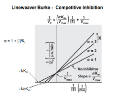
  • Competitive inhibitors- the inhibitor and the substrate resemble each other; the inhibitor binds to the active site of the enzyme; Km will increase whereas the Vmax will not change.
  • Uncompetitive inhibitors- the inhibitor does not resemble the substrate, the inhibitor binds only to the substrate-enzyme complexKm and Vmax decreases to the same amount.
  • Non-competitive inhibitors- is a form of mixed inhibition; the inhibitor does not resemble the substrateVmax decrease, Km is unaffected.
  • Mixed inhibitors – the inhibitor can bind to the enzyme at similar times as the enzyme’s substrate; Vmax decreases, while Km may either increase OR decrease.

The Structures of Proteins

proteins

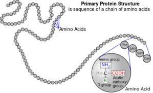
Proteins are made up of amino acids which undergo condensation reactions to form covalent peptide bonds.

Proteins can have different levels of structure. Hundreds or thousands of amino acids undergo condensation reactions in a particular sequence to form a polypeptide chain which can then fold or bend in different ways at which point it will be called a protein.

The primary structure of a protein refers to the number of amino acids found in the protein, that is their sequence and the number of polypeptide chains making up the protein.

1'

The secondary structure of proteins:

All proteins have a secondary structure which is due to the hydrogen bonding between the CO of 1 amino acid and the NH of another amino acid. This results in the formation of an α- helix which is maintained by the hydrogen bonds. The secondary structure of a protein may also be in the form of β-pleated sheets. This structure is the result of the hydrogen bonding which occurs between the polypeptide chains. The C=O of 1 amino acid on a polypeptide chain undergoes hydrogen bonding with the hydrogen of the NH group on another, parallel polypeptide chain. A protein like this has high tensile strength and is flexible (easily folded) due to its sheet-like form which is produced due to the hydrogen bonds. This protein does not stretch because of the intensity of the hydrogen bonds holding the polypeptide chains together.

2'

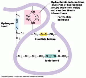
The tertiary structure of a protein is the result of the hydrogen bonding between strongly polar R-groups, ionic bonding between the ionized amino acids and the carboxylic groups, hydrophobic interactions between non-polar side chains and disulphide bridging between cysteine molecules. The hydrogen bonding may also occur between the NH groups of the polypeptide chain. In this level of protein structure, the secondary structure is folded on itself because of the various types of bonding to form a very compact molecule. It is usually folded in such a way that the hydrophilic R-groups are on the outside of the folded protein and the hydrophobic R-groups lie in the interior of the folded protein. Such proteins dissolve because of the fact that hydrogen bonding can occur in water.

3'

The quarternary structure of a protein is due to more than one polypeptide chain being held together by hydrogen bonding, hydrophobic interactions and ionic bonding. A protein which shows this type of structure is haemoglobin which consists of four polypeptide chains. Some proteins can also have a non-polypeptide structure called a prosthetic group. These proteins are called conjugated proteins. The haem group in haemoglobin is a prosthetic group.

 4'